
Pvt Rhythm Strip
(Cease bolus’ at indication of fluid in lungs showing repiratory distress or rales.
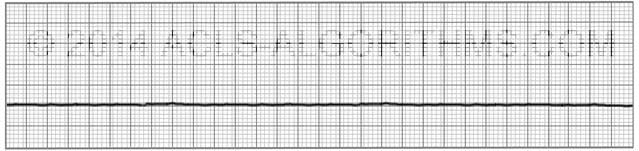
Pvt rhythm strip. Asystole is a flat-line ECG (Figure 27). It will pull up a page with an example strip and an easy to understand deicription. How Your Heart Beats Your heart is a muscular organ that pumps about 100,000 times a day to.
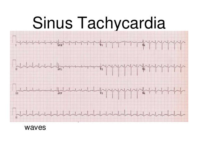
Electrocardiogram (ECG) shows regular R waves in the absence of atrial rhythm. For many people, treatment and lifestyle changes can control or eliminate this heart rhythm problem. A premature contraction feels like your heart "skipped a beat." Premature atrial contractions (PACs) start in the upper chambers of the heart (atria).
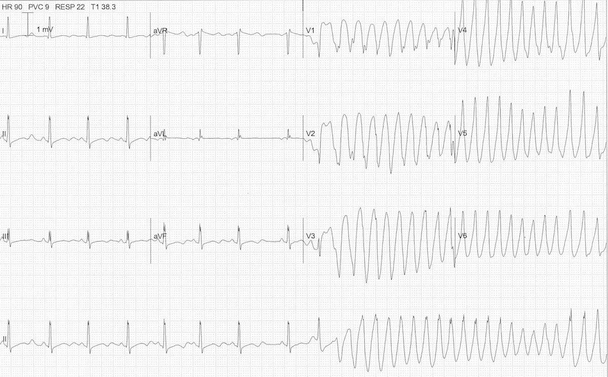
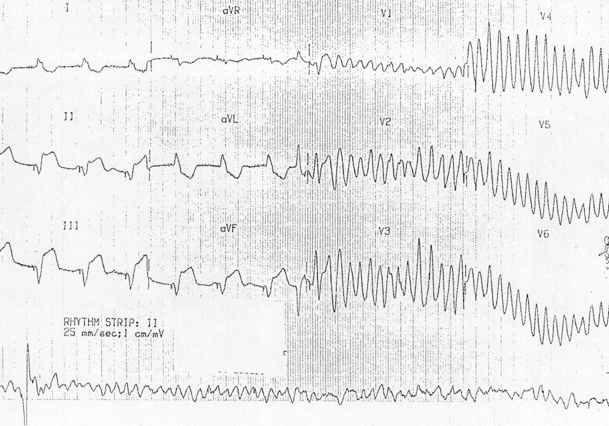
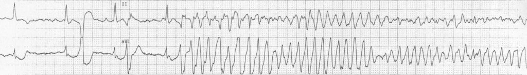
The atrial fibrillation is interrupted by a rapid and regular tachycardia with wide QRS complex. Suitable for beginner, intermediate, advanced & expert pole dancers ***GET 10% OFF OPEN DANCE ACADEMY***. Assess rhythm and possible cause;.
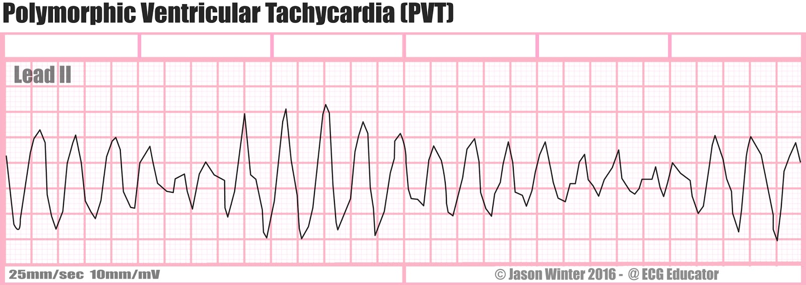
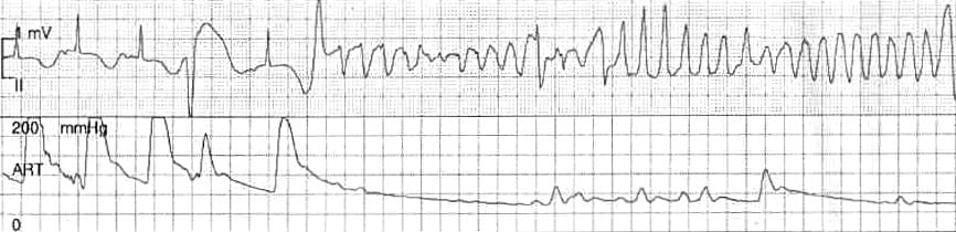
The ventricles suddenly attempt to contract at rates of up to 500 bpm. Navigate to our home page to learn more!. Qc サークル イラスト~pvt rhythm strip 三現主義 現場 現物 現実 を定着化する 製造業 品質改善の進め方 Qc サークル イラスト.
Users are challenged to identify each type of arrhythmia based upon the cardiac rhythm strip. Pediatric Learning Solution's Augmented Reality Polymorphic Ventricular Tachycardia (PVT) job aid provides an overview of PVT and an animated ECG display of the rhythm and its effect on the beating heart. After opening or printing out the job aid, tap the "Augmented Reality" button in the PLS App to start the AR viewer.
These cards can be used in a variety of classroom activities, such as whole class instruction, music centers, or cooperative learning. Premature ventricular contractions (PVCs) are extra heartbeats that begin in one of your heart's two lower pumping chambers (ventricles). Ventricular tachycardia is an abnormal ventricular rhythm with a pulse rate above 100 beats per minute.
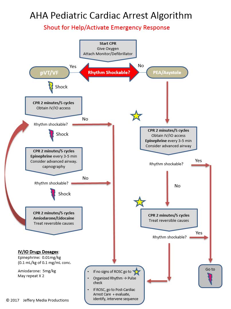
Any organized rhythm without detectable pulse is “PEA” ACLS Rhythms for the ACLS Algorithms 255 3. After initiating CPR and assessing the rhythm, the patient is in VF/ pVT and the first shock is given, and CPR for 2 mins.Then rhythm check and in VT, should the pulse be checked to confirm pVT?. Clinically accurate heart rate monitoring giving doctors uninterrupted ECG recordings.
Although a few seconds may not result in problems, longer periods are dangerous. Blood gas, lactate, glucose, CBC, ionized calcium, cultures. Cardiac Rhythms Strips and Drills.
Also, cease bolus’ if hepatomegaly presents.). Iqwal Mangat, Paul Dorian, in Clinical Critical Care Medicine, 06. They're the most common reason for.
Rhythm Strip Flash Card Practice Click or tap the cards to see the answer. The P Wave is the usual first waveform component and indicates electrical activity that triggers atrial contraction. Quickly find any rhythm and click go.
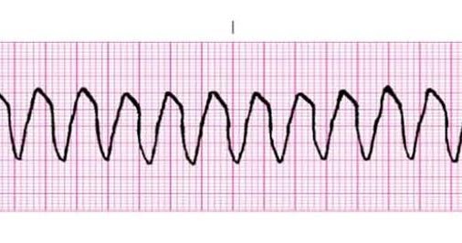
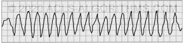
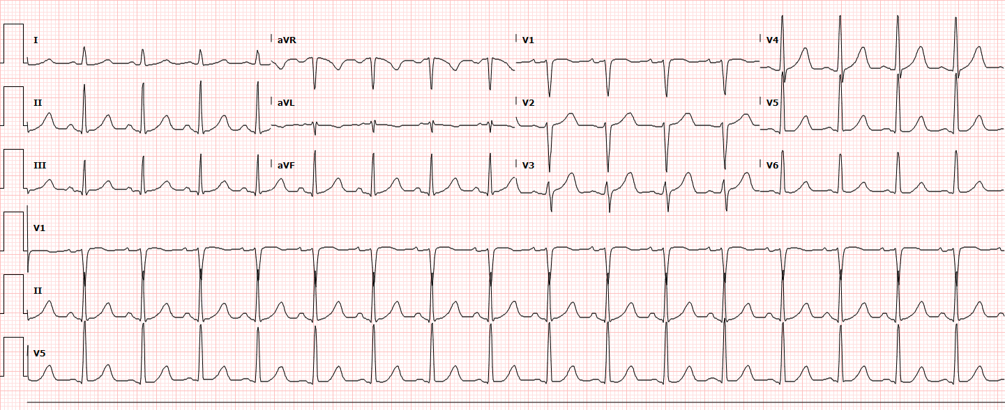
Monomorphic Ventricular Tachycardia (Wide and Regular) Monomorphic ventricular tachycardia (MVT) can occur in various settings, most importantly in the setting of structural heart disease. QRS = duration (0.06 - 0.10 ), voltage, q or Q waves 6. Also with V-tach because if its untreated it may lead to V-Fib.
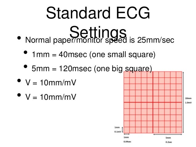
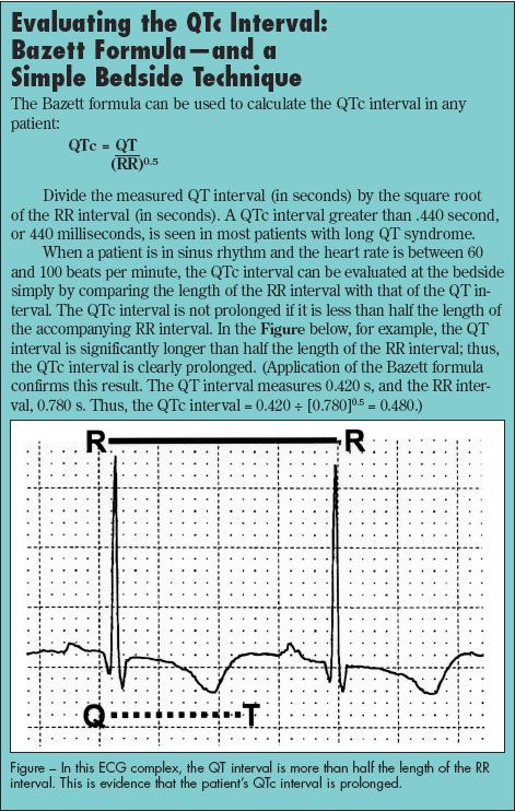
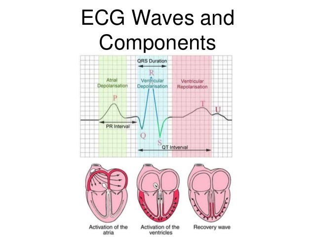
P-R interval = length (0.12 - 0.2 sec = <1 big square), isoelectric. Multiple electrical events called P, Q, R, S, T and U may be observed on an EKG strip. No lengthy deep learning.
The amiodarone was not to use amiodarone as an actual treatment modality as an isolated means to treat the “non sinus rhythm. Wolff-Parkinson-White syndrome (WPW) is the most common type of PSVT in. Most important will be definitely V-Fib and Asystole.
ACLS Cardiac Arrest VTach and VFib Algorithm Perform the initial assessment Perform high-quality CPR Establish an airway and provide oxygen to keep oxygen saturation > 94% Monitor the victim’s heart rhythm and blood pressure If the patient is in VTach or VFib, this IS a shockable rhythm Apply defibrillator pads (or paddles) and shock the …. Heart Block Tricks from Terry 4 RHYTHM & ♥ ETIOLOGY CRITERIA OVERVIEW UNIQUE CRITERIA SAMPLE STRIPS ***2nd degree AV Block Type II or Mobitz II RHY – Regular or Irregular PRI – constant QRS – normal or wide Regular or Irregular EXTRA Ps 3rd degree AV Block. Has over 450 online lessons, by some world famous pole dancing champions!.
They can be used to reinforce rhythms students are learning, both vocally and on various instruments. This rapid and irregular electrical activity renders the ventricles unable to contract in a synchronised manner, resulting in immediate loss of cardiac output. Map P-P and R-R intervals.
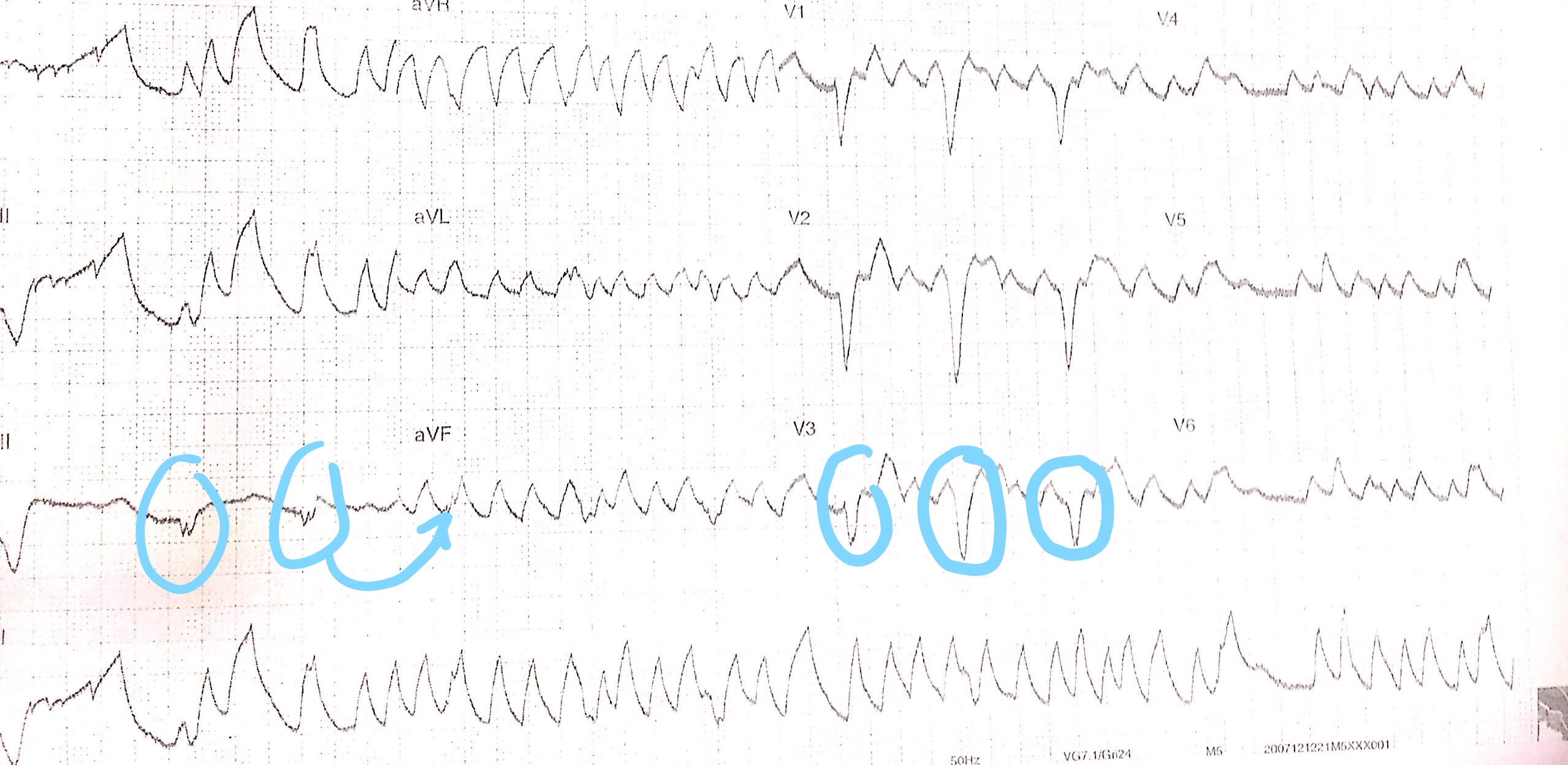
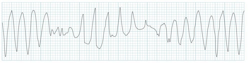
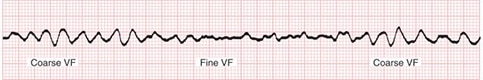
The two images show what ventricular fibrillation will look like on an EKG rhythm strip. ST Segment = shape, isoelectric with PR segment. All R waves are.
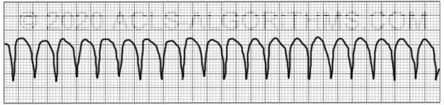
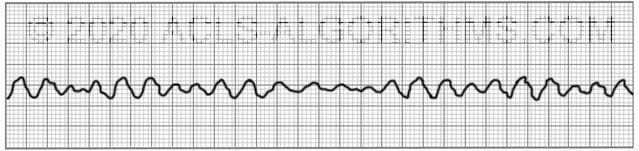
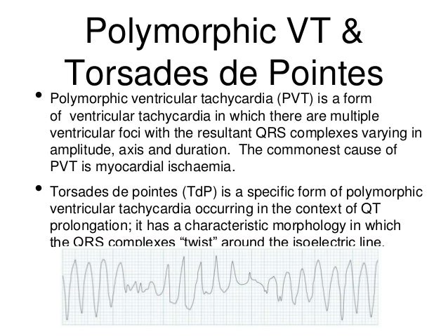
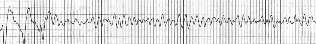
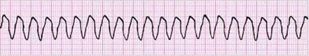
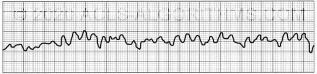
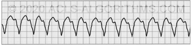
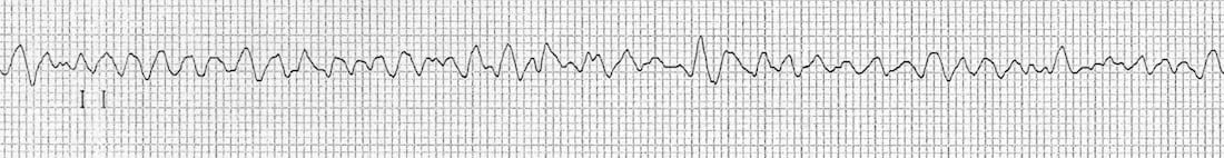
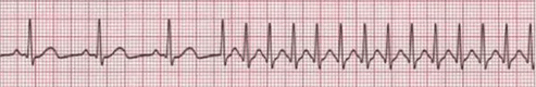
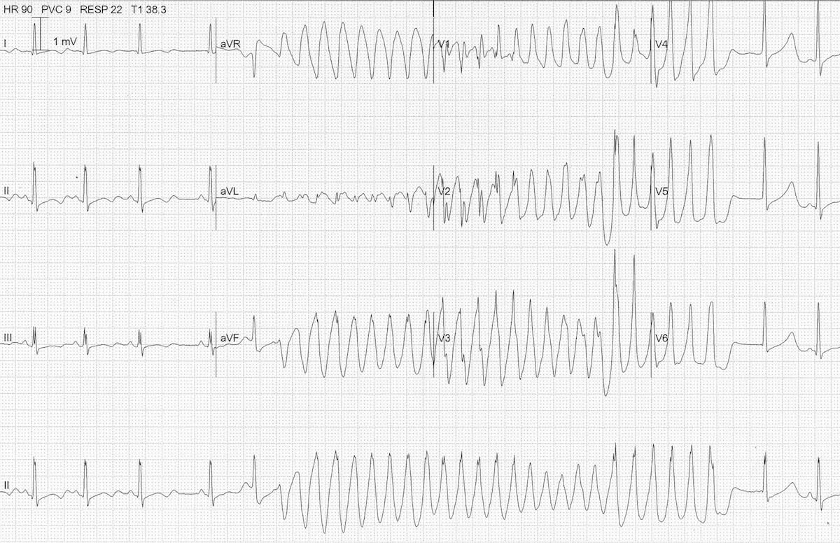
This rhythm usually appears on the monitor as a wide, regular, and very rapid rhythm. Torsades de pointes is commonly recurrent. It is the most frequent abnormal heart rhythm in newborns and infants.
Just find your strip fast and easy!. If your heart feels out of rhythm or "flutters," especially when you have a lot of anxiety, it could be caused by premature ventricular contractions, or PVCs. Analyze the rhythm strip:.
Ventricular tachycardia (v-tach) typically responds well to defibrillation. Because of the preceding long QU interval, this can be diagnosed as TdP. There may be subtle movement away from baseline (drifting flat-line), but there is no perceptible cardiac electrical activity.
Short periods may occur without symptoms, or present with lightheadedness, palpitations, or chest pain. Hone your skills as an accomplished interpreter of 12-lead ECGs and rhythm strips. PR Interval PR interval is not measured since this is a ventricular rhythm.
Description A rapid heart rate can originate in either the left or right ventricle. Come join us in the Advanced ECG Interpretation Boot Camp or the Masterclass in Advanced Electrocardiography. These extra beats disrupt your regular heart rhythm, sometimes causing you to feel a fluttering or a skipped beat in your chest.
Ventricular fibrillation (VF) is the the most important shockable cardiac arrest rhythm. As seen in the beginning of the recording, the patient has an underlying rhythm of atrial fibrillation. If there is no rhythm change and the same waveform of VT continues, you would not need to perform a pulse check.Performing the pulse check would delay the continuation of chest compressions.
Determine rate and regularity of paced activity. AV sequential pacing with appropriate sensing and capture. Rhythm Strip Samples to help with ACLS Precourse Assessment with Unique Criteria.
Analyze the strip for failure to capture, failure to sense, oversensing or failure to pace. To be classified as tachycardia, the heart rate is usually at least 100 beats per minute. The initiation or continuation of lidocaine may be considered immediately after ROSC from cardiac arrest due to VF/pVT.
Search for an EKG strip from a simple drop down list. On the ECG rhythm strip, a ventricular premature beat followed by a long pause followed by a conducted QRS complex and a ventricular premature beat, which initiates the arrhythmia, is commonly seen (the so-called long-short initiation;. Read on to learn about the causes, symptoms, and more.
Accessed November 27, 18. Comment on the underlying rhythm if it is determinable. Supraventricular tachycardia (SVT) is an abnormally fast heartbeat that originates above the ventricles in the atria or AV node.
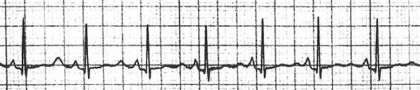
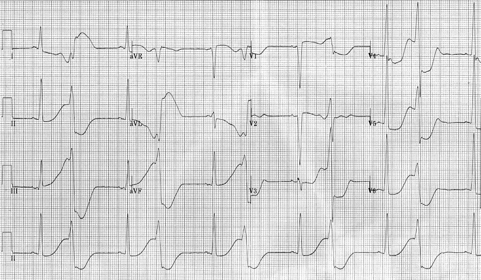
A premature atrial complex (beat #9 of the rhythm strip) lands on the end of the T wave, causing ‘R on T’ phenomenon and initiating a paroxysm of polymorphic VT. Example 3 TdP secondary to hypokalaemia:. Paroxysmal atrial tachycardia is a type of irregular heartbeat that increases heart rate.
Stay with me here…. Join now to get:. Definition Ventricular tachycardia (V-tach) is a rapid heart beat that originates in one of the lower chambers (the ventricles) of the heart.
I have even included a rhythm practice strips with answers and explanation. Your doctor can try to bring your heart back into a regular rhythm with medicines and other treatments. 1 Sinus Brady - Arrhythmia The rate is slow and the rhythm is irregular 2 Sinus Brady - heart rate is less than 60 3 Normal Sinus Rhythm 4 Normal Sinus Rhythm 5.
Excellent value for money when compared to the price of local pole dancing classes;. Ventricular tachycardia presents with palpitations, chest pain, and difficulty in breathing. Access to more than 450 high-quality video lessons with the lifetime access option ($350);.
They may also come in a state of cardiac arrest. The Zio Patch is a complete ambulatory cardiac monitoring solution. Arrhythmia is an irregular cardiac rhythm or an abnormally fast or slow heart beat.
Interpreting EKG Rhythm Strips Step 2 – Rhythm Measuring a Regular Rhythm Measure the intervals between R waves (measure from R to R) If the intervals vary by less than 0.06 seconds or 1.5 small boxes, the rhythm is considered to be regular. The 4th beat from the end is a premature ventricular beat and its QRS morphology is identical to the QRS seen during the tachycardia. These are the Pulseless rhythm that you need to watch out for.
These Rhythm Strips Beginning Rhythms are great for use in any music classroom or private music studio. Paroxysmal atrial tachycardia (PAT) is a condition in which the upper chambers of the heart, the atria, begin to beat irregularly, sometimes producing heartbeats as fast as 0-2 beats per minute (bpm).The condition is called paroxysmal, since it occurs suddenly and without warning.In people with no abnormal heart conditions, this is usually considered not a dangerous arrhythmia, but in. The ECG criteria to diagnose premature ventricular contractions (PVCs) is discussed with 12-lead ECG examples including ventricular bigeminy and ventricular trigeminy.
Ventricular tachycardia refers to a wide QRS complex heart rhythm — that is, a QRS duration beyond 1 milliseconds — originating in the ventricles at a rate of greater than 100 beats per minute. Fast (>100 beats/min) or slow. There are 2 ways of classifying EKG rhythms making it a whole lot easier to interpret any rhythm presented to you, be it in clinical setting, in class, ACLS or in an EKG Rhythm Test.
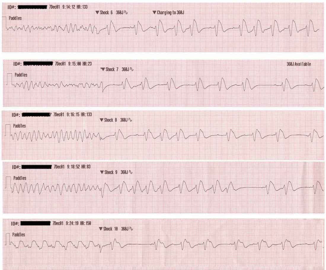
University of Virginia School of Medicine. Cardiac Rhythm Strip Drill. If a shockable rhythm is present, either v-fib or pulseless v-tach, begin the charging sequence on the defibrillator and resume chest compressions until the defibrillator is charged.
Premature ventricular contractions (PVCs) start in the lower chambers of the heart (ventricles). Most NCLEX questions will show you the rhythm strips and you have to know what it is and it will all you what will the nurse do first or best etc. PEA (Pulseless Electrical Activity) Defining Criteria per ECG Rhythm displays organized electrical activity (not VF/pulseless VT) Seldom as organized as normal sinus rhythm Can be narrow (QRS <0.10 mm) or wide (QRS >0.12 mm);.
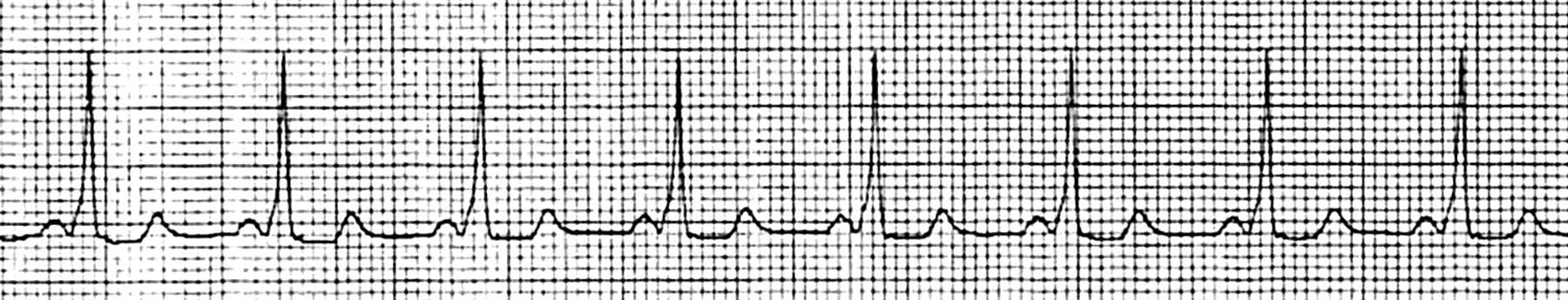
There are occasionally P waves in the strip, but they are not associated with the ventricular rhythm. Provided repeated IV bolus’ of crystalloids at ml/kg. PSVT affects about 1 in every 2,500 children.
In these patients, MVT occurs as a result of a reentrant mechanism, usually because of areas of scar interspersed with areas of slow. Here are the 2 ways to classify EKG Rhythms;. Rhythm = Regular or irregular.
After each answer, immediate feedback and coaching is available. Pulseless electrical activity (PEA) and asystole are related cardiac rhythms in that they are both life-threatening and unshockable. Premature ventricular contractions are common — they occur in many people.
Ventricular tachycardia may result in ventricular. P wave = present, 1 per QRS h d ti ltQRS, shape, duration, voltage. Ventricular tachycardia (V-tach or VT) is a type of regular, fast heart rate that arises from improper electrical activity in the ventricles of the heart.

How To Interpret Read Ekgs Like A Boss Master Heart Rhythms Education Nursejanx
Polymorphic Vt And Torsades De Pointes Tdp Litfl

Ecg Week Of 5 08 Interpretation Emergucate
Pvt Rhythm Strip のギャラリー
Www Emnote Org Uploads 1 3 5 3 Arrhythmia Pdf
Med Virginia Edu Emergency Medicine Wp Content Uploads Sites 232 15 10 Introduction To Ecg Rhythms Pdf
2

R From T As A Common Mechanism Of Arrhythmia Initiation In Long Qt Syndromes Circulation Arrhythmia And Electrophysiology

Syncope In A Woman With A History Of Myocardial Infarction Consultant360
Ventricular Tachycardia After Ondansetron Administration In A Child With Undiagnosed Long Qt Syndrome Springerlink
Ecg Analysis
Ventricular Fibrillation Acls Algorithms Com

R Review Cardio Conduction D O Flashcards Quizlet
Med Virginia Edu Emergency Medicine Wp Content Uploads Sites 232 15 10 Introduction To Ecg Rhythms Pdf

Normal To Advanced Rhythms Springer Publishing
2
Dr Smith S Ecg Blog Polymorphic Ventricular Tachycardia
Syncope In A Woman With A History Of Myocardial Infarction
Ventricular Fibrillation Vf Litfl Ecg Library Diagnosis

A Interrogation Strip From Pacemaker Showing An Atrial Sensed Download Scientific Diagram

Ecg Educator Blog 11 14 16

A Initial 12 Lead Electrocardiogram For Patient Described In Case 1 Download Scientific Diagram
1
Http Www Lbstack Com Students Syncope Syncope Pdf

Ventricular Arrhythmias Basic And Bedside Electrocardiography 1st Edition 09
Q Tbn 3aand9gct36gy8ntg2rnsquokblhaenwwy6vgidc8ki5pv87zwpfzzdbxo Usqp Cau
2
Ecg Week Of 5 08 Interpretation Emergucate

Acls Ventricular Fibrillation And Pulseless Ventricular Tachycardia Guide
Med Virginia Edu Emergency Medicine Wp Content Uploads Sites 232 15 10 Introduction To Ecg Rhythms Pdf
Http Www Nwcemss Org Assets 1 System Entry Ventricular Rhythms F17 Pdf
Other Tachycardia Rhythms Acls Algorithms Com
Polymorphic Vt And Torsades De Pointes Tdp Litfl
Pulseless Ventricular Tachycardia Acls Algorithms Com

Effects Of Left Ventricular Assist Device Therapy On Ventricular Arrhythmias Sciencedirect
Polymorphic Ventricular Tachycardia Pvt Is A Form Of Ventricular Tachycardia In Which
Ecg Analysis

Ventricular Arrhythmias Basic And Bedside Electrocardiography 1st Edition 09
Sw Ohio Pulseless Rhythms Ekg For Asystole Pea Vt Vf Flashcards Memorang

R From T As A Common Mechanism Of Arrhythmia Initiation In Long Qt Syndromes Circulation Arrhythmia And Electrophysiology
Med Virginia Edu Emergency Medicine Wp Content Uploads Sites 232 15 10 Introduction To Ecg Rhythms Pdf
Q Tbn 3aand9gcqts24zy8 O4ata1y1l95dt16bmfklbcgfih0gkrhxza2ulzs Y Usqp Cau

A Resting 12 Lead Ecg On Admission B Rhythm Strip Showing Transient Download Scientific Diagram
Ecg Analysis
Electrocardiographic Features Of Wolff Parkinson White Syndrome Emergency Medicine Journal

Ventricular Tachycardia V Tach Or Vt Is A Type Of Regular And Fast

How To Interpret Read Ekgs Like A Boss Master Heart Rhythms Education Nursejanx
Http Heartcentertraining Com Wp Content Uploads 13 11 Precourseassessment Pdf
Http Www Resuscitationjournal Com Article S0300 9572 14 1 Pdf
2

Dr Smith S Ecg Blog Polymorphic Ventricular Tachycardia
Med Virginia Edu Emergency Medicine Wp Content Uploads Sites 232 15 10 Introduction To Ecg Rhythms Pdf
Ecg Analysis

Dr Smith S Ecg Blog Polymorphic Ventricular Tachycardia
Ventricular Fibrillation Vf Litfl Ecg Library Diagnosis

Ventricular Arrhythmias Basic And Bedside Electrocardiography 1st Edition 09

Pharmacology In Cardiac Arrest
Http Www Nwcemss Org Assets 1 System Entry Ventricular Rhythms F17 Pdf
Www Emnote Org Uploads 1 3 5 3 Arrhythmia Pdf
Gale Onefile Health And Medicine Document Country Cardiograms Case 31

Shockable Rhythms Ventricular Tachycardia Ventricular Fibrillation Supraventricular Tachycardia Acls Com

Ventricular Fibrillation Wikipedia
Ventricular Fibrillation Vf Litfl Ecg Library Diagnosis

Normal To Advanced Rhythms Springer Publishing
Www Emnote Org Uploads 1 3 5 3 Arrhythmia Pdf
1

A 12 Lead Electrocardiogram For Patient Described In Case 2 B Download Scientific Diagram
Med Virginia Edu Emergency Medicine Wp Content Uploads Sites 232 15 10 Introduction To Ecg Rhythms Pdf
Matters Of The Heart V Tach Patmac Rn

Polymorphic Ventricular Tachycardia An Overview Sciencedirect Topics

Make It Two A Case Report Of Dual Sequential External Defibrillation
Polymorphic Vt And Torsades De Pointes Tdp Litfl
Ventricular Fibrillation Acls Algorithms Com

Acls Ventricular Fibrillation And Pulseless Ventricular Tachycardia Guide

Ventricular Arrhythmias Basic And Bedside Electrocardiography 1st Edition 09

Ventricular Fibrillation 네이버 블로그

Im Crazy About Ecg Home Facebook
2
Shockable Rhythms Ventricular Tachycardia Ventricular Fibrillation Supraventricular Tachycardia Acls Com

Examples Of The Occurrences Of Sve Mvt And Pvt Classification Was Download Scientific Diagram
Shockable Rhythms Ventricular Tachycardia Ventricular Fibrillation Supraventricular Tachycardia Acls Com

A Initial Pulseless Ventricular Tachycardia Rhythm Strip B Download Scientific Diagram
Http Www Nwcemss Org Assets 1 System Entry Ventricular Rhythms F17 Pdf

Ekg Rhythym Emergency Nursing Icu Nursing Critical Care Nursing

Acls Ventricular Fibrillation And Pulseless Ventricular Tachycardia Guide
Ventricular Fibrillation Acls Algorithms Com

Matters Of The Heart V Tach Patmac Rn
Polymorphic Vt And Torsades De Pointes Tdp Litfl
Other Tachycardia Rhythms Acls Algorithms Com
Ventricular Fibrillation Vf Litfl Ecg Library Diagnosis
Shockable Rhythms Ventricular Tachycardia Ventricular Fibrillation Supraventricular Tachycardia Acls Com
Lms Rn Com Getpdf Php 633 Pdf
Polymorphic Vt And Torsades De Pointes Tdp Litfl
Electrocardiography Chapter 10 Worksheet
Lms Rn Com Getpdf Php 633 Pdf
Ventricular Tachycardia s Differential Diagnosis Of

Advanced Life Support 1 2 Flashcards Quizlet

Ecg Educator Blog Six Second Ecg Rhythm Strips

Supraventricular Tachycardia Wikipedia

How To Interpret Read Ekgs Like A Boss Master Heart Rhythms Education Nursejanx
Polymorphic Vt And Torsades De Pointes Tdp Litfl
2

Acls Pulseless Electrical Activity And Asystole Guide
A Missense Mutation In A Highly Conserved Region Of Casq2 Is Associated With Autosomal Recessive Catecholamine Induced Polymorphic Ventricular Tachycardia Ppt Download

47 Year Old Male Cc Crushing Chest Pain Conclusion Ems 12 Lead


